| Basil Diseases | |||||||||||||||||
|---|---|---|---|---|---|---|---|---|---|---|---|---|---|---|---|---|---|
|
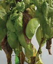
Back to Basil Page 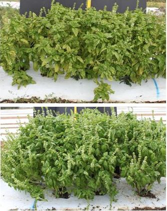 Fig. 1 Symptoms of downy mildew on field-grown basil (top) compared with healthy basil plants (bottom) 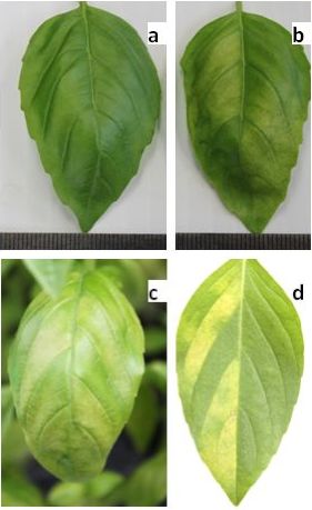 Fig. 2 Top view of basil leaf a) without any disease and b-d) exhibiting chlorosis and vein-bounded yellowing of downy mildew 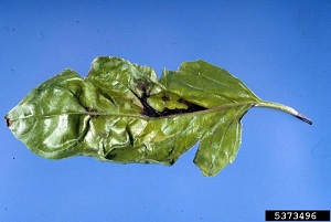 Fig. 6  Basil bacterial leaf spot sign Pseudomonas cichorii  Fig. 9  Basil with fusarium wilt. Notice wilted, yellow leaves and distorted new growth Back to Basil Page |
Downy
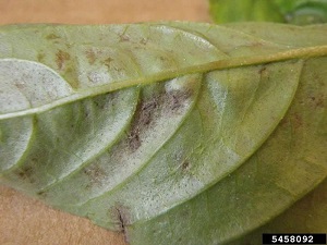
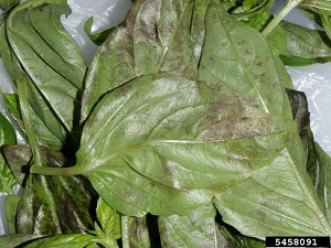
Mildew Caused by Peronospora belbahrii Symptoms initially appear as yellowing of leaves and are typically concentrated around the middle vein (Fig. 2). The discolored area may cover most of the leaf surface. On the underside of leaves, a gray fuzzy growth of the pathogen may be apparent by visual inspection (Fig. 3). Under high humidity, the chlorotic areas turn to dark brown quickly. Sporangia, the reproductive structures of the pathogen, are easily detected under magnification. 1 All sweet varieties are very susceptible to downy mildew. Red types, Thai basil, lemon basil, lime basil, and other spicy basils have been found to be less susceptible. 5 A few fungicides are currently labeled for downy mildew control on basil. Some phosphorous acid fungicides are effective against downy mildew under herbs on the current label. These fungicides were effective in fungicide efficacy experiments with applications started before or after initial symptoms were found. 2 Young basil plants are more susceptible than old basil plants (Patel et al. 2014), and therefore plants should be protected starting from their emergence from the ground. Reducing the period of leaf wetness by avoiding overhead watering may also be helpful. Heavily infected plants should be discarded. If possible, isolate new plantings to reduce inoculum spread from older plantings. 2
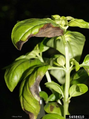
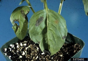
Fig. 3. A closer view of a lesion with dense sporulation that looks like a grayish-purple mold on the leaf Fig. 4. Heavy spore production on the undersurface of the leaf may look like dust or dirt on the tissue Fig. 5. Basil downy mildew symptoms Peronospora belbahrii Further Reading Downy Mildew of Basil in South Florida, University of Florida pdf Leaf Spot Caused by Colleotrichum sp. Dark spots form on leaves and the dead tissue within leaf spots may drop out, causing a shot-hole symptom. The disease can cause defoliation, tip dieback, stem lesions, and sometimes loss of entire plants. Spores are water-splashed from diseased tissue. 1 Sow seed in sterile containers in sterilized soil or a soilless mix. Since wet conditions favor disease development, reduce periods of leaf wetness by reducing humidity and increase plant spacing to increase air movement. Avoid overhead irrigation. Remove diseased plants to reduce inoculum levels. 1 Bacterial Leaf Spot Caused by Pseudomonas cichorii Spots on leaves are water-soaked and dark. They may be both angular and delineated by small veins in the leaves or irregular in shape. A wet stem rot may occur. Bacterium is reported to be seed-borne. The disease is favored by wet, humid conditions and is disseminated by splashing water or by handling infected tissue and then touching other plants. 1 Decrease moisture on plants with low humidity and sufficient plant spacing for adequate movement to reduce periods of leaf wetness. Use disease-free seed and transplants. Remove diseased leaves and plants to reduce inoculum levels. Avoid overhead irrigation. Use clean, sterile equipment and do not move between infected and healthy plants. 1
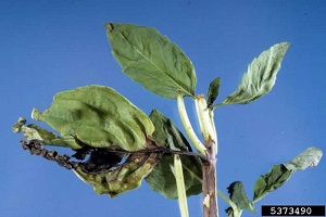
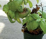
Fig. 7,8. Basil bacterial leaf spot sign. Pseudomonas cichorii Fusarium Wilt Caused by Fusarium oxysporum Early symptoms of fusarium wilt disease are yellowed shoots, twisted young leaves, internal discoloration within the stem, and a few rotted leaves. As the disease advances, a black stem rot progresses upward, the plants wilt, the shoots die back, the leaves drop, and the plant may die. 3 Fusarium wilt of basil is caused by a soil-borne fungus (Fusarium oxysporum f. sp. basilicum). The fungus attacks the water-conducting tissue (xylem) within the stem. Infected basil plants will grow normally until they are six to twelve inches tall, then become stunted and exhibit browning of terminal growth. Once water uptake is totally blocked, the basil plants will suddenly wilt. Promptly remove and dispose of any wilt-infected plants. 4 The Fusarium wilt pathogen can survive for many years in the soil; therefore plant resistant basil varieties. Three Genovese-type cultivars of basil have been selected for resistance to Fusarium wilt. The first cultivar released was ‘Nufar’, which grows to 24 inches tall and has medium-sized leaves with mild flavor. More recently ‘Aroma 1’ and ‘Aroma 2’ have been released and both have very good fragrance. These cultivars are readily available from mail-order seed companies. 4 Use disease-free seed in sterilized soil or a soilless mix. Seed may be disinfested. Rotate fields to another crop besides basil. 1
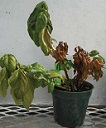
Fig. 10. Symptoms of basil fusarium wilt including stem-lesions (often one-sided initially), wilting and leaf yellowing. As symptoms progress, older leaves may fall off plant Fig. 11. Stems can become distorted and exhibit a characteristic "shepherd's crook" as seen in the dead stem in this picture Fig. 12. Fusarium wilt infection of three basil plants in the same container. Notice plants on right were infected first and eventually the disease progressed to plant on the left Further Reading Basil Fusarium Wilt, e-GRO Alert pdf Fusarium Wilt Of Basil, University of Illinois Extension pdf 'UH sweet Basil'. A New Basil Cultivar Tolerant of Fusarium Wilt, University of Hawai'i, Cooperative Extension pdf Further Reading Florida Plant Disease Management Guide: Sweet Basil, University of Florida pdf |
||||||||||||||||
| Bibliography 1 Zhang, Shouan, and Pamela D. Roberts. "Florida Plant Disease Management Guide: Sweet Basil." Plant Pathology Dept., UF/IFAS Extension, PP-113, Original pub. Jan. 2003, Revised Mar. 2009 and Dec. 2021, AskIFAS, edis.ifas.ufl.edu/pp113. Accessed 21 Dec. 2017, 20 Dec. 2023. 2 Zhang, Shouan, et al. "Downy Mildew of Basil in South Florida." Plant Pathology Dept., UF/IFAS Extension, PP271, Original pub. Sept. 2009, Revised Dec. 2015, AskIFAS, edis.ifas.ufl.edu/pp271. Accessed 21 Dec. 2017. 3 Hamasaki, Randall T. "'UH Sweet Basil' A New Basil Cultivar Tolerant of Fusarium Wilt." University of Hawaii, 1 p. (New Plants for Hawaii; NPH-3), May 1998, /hdl.handle.net/10125/12183. Accessed 20 Mar. 2014. 4 Williamson, Joey. "Basil." Clemson University Cooperative Extension, South Carolina, Dec. 2008, www.clemson.edu/extension. Accessed Mar. 2014. 5 "Basil." Gardening Solutions, University of Florida,AskIFAS, gardeningsolutions.ifas.ufl.edu/plants/edibles/vegetables/basil.html. Accessed 6 Oct. 2018. Photographs Fig. 1 Mersha, Zelalem. Symptoms of downy mildew on field-grown basil (top) compared with healthy basil plants (bottom). edis.ifas.ufl.edu/LyraEDISServlet?command=getImageDetail&image_soid=FIGURE%201&document_soid=PP271&document_ version=7119. Accessed 21 Dec. 2017. Fig. 2 Mersha, Zelalem. Top view of basil leaf a) without any disease and b-d) exhibiting chlorosis and vein-bounded yellowing of downy mildew. edis.ifas.ufl.edu//LyraEDISServlet?command=getImageDetail&image_soid=FIGURE 2&document_soid=PP271&document_version=7119. Accessed 21 Dec. 2017. Fig. 3 Jensen, Sandra. "5458092. Basil Downy Mildew Peronospora belbahrii Sign." 31 Jan. 2012. Cornell University. Bugwood.org. (CC BY-NC 3.0 US), www.forestryimages.org/browse/detail.cfm?imgnum=5458092. Accessed 21 Mar. 2014. Fig. 4 Jensen, Sandra. Heavy spore production on the undersurface of the leaf may look like dust or dirt on the tissue. Uploaded January 31, 2012, Updated February 3, 2012. (CC BY-NC 3.0 US), www.forestryimages.org/browse/detail.cfm?imgnum=5458091. Accessed 21 Mar. 2014. Fig. 5 Watt, Bruce. Basil downy mildew (Peronospora belbahrii). University of Maine, Bugwood.org. 5458610. 2012. (CC BY-NC 3.0 US), www.forestryimages.org/browse/detail.cfm?imgnum=5458610. Accessed 21 Mar. 2014. Fig. 6 Bacterial leaf spot (Pseudomonas cichorii) symptoms. Florida Division of Plant Industry, Florida Department of Agriculture and Consumer Services, Bugwood.org. Uploaded June 24, 2008, Updated September 8, 2008. (CC BY-NC 3.0 US), www.forestryimages.org /ipmimages.org/browse/detail.cfm?imgnum=5373496. Accessed 21 Mar. 2014. Fig. 7 Bacterial Leaf Spot Pseudomonas cichorii Sign. 2008. Florida Division of Plant Industry Archive, Florida Department of Agriculture and Consumer Services. Bugwood.org. (CC BY-NC 3.0 US), www.forestryimages.org/detail.cfm?imgnum=5373490. Accessed 21 Mar. 2014. Fig. 8 Bacterial Leaf Spot Pseudomonas cichorii Sign. 2008. Florida Division of Plant Industry Archive, Florida Department of Agriculture and Consumer Services. Bugwood.org. (CC BY-NC 3.0 US), www.forestryimages.org/browse/detail.cfm?imgnum=5373492. Accessed 21 Mar. 2014. Fig. 9,10,11,12 Basil with fusarium wilt. Basil Fusarium Wilt. e-Grow Edible Alert, Vol. 1, No. 2, Jan. 2016, www.e-gro.org/pdf/E102.pdf. Accessed 21 Dec. 2017. Published Mar. 2014 KJ. Last update 20 Dec. 2023 LR |
|||||||||||||||||
